Body Skin · Research Explainer
Why the Skin on Your Elbows Becomes Crepey After 40
The elbow develops crepey skin in two structurally distinct zones that age through different mechanisms. Understanding the biology of each — and what they share at the molecular level — explains why the elbow is one of the most reliably affected sites on the arm in midlife.

When women describe crepey skin at the elbows, they tend to describe two distinct phenomena. The first is the accordion-fold at the inner crease — the antecubital fossa — where the skin bunches into a permanent crinkle even when the arm is extended, and where a deep, paper-thin fold appears with every bend. The second is the outer elbow cap, the olecranon region, where the skin looks ashy, tissue-thin, and rough in a way that has nothing to do with dryness and does not respond to moisturiser.
Both zones are on the same joint, and both reflect crepey texture. But the structural reasons they arrive at crepey texture are not identical. Understanding each zone separately — and what they share at the molecular level — is essential to understanding why the elbow develops this pattern so reliably after 40.
Zone one: the antecubital fossa and the mechanics of extreme flexion
The inner elbow crease — the antecubital fossa — is the hinge point of one of the most used joints in daily life. Every time you reach for something, type at a keyboard, lift a glass, or cradle a phone between ear and shoulder, the skin at the inner elbow folds, stretches, and unfolds. The angle of flexion at the elbow is steeper and more complete than at the knee, and the frequency of movement is substantially higher. The inner elbow crease participates in hundreds of deformation cycles per day from early adulthood onward.
Skin is viscoelastic. It deforms under load and, in younger dermis with intact collagen and elastin fibres, it recovers its resting geometry after the deformation is released. This recovery depends on the architectural integrity of the dermal scaffold — the cross-linked network of collagen type I and III fibres woven through an elastin fibre mesh, embedded in a hydrating matrix of hyaluronic acid and proteoglycans. When the scaffold is intact and well-maintained, the skin at the antecubital fossa handles extreme flexion cycles and returns cleanly to shape.
The problem is that collagen synthesis declines at approximately one percent per year from the mid-twenties onward. The collagen in the inner elbow crease is being mechanically fatigued at a high cycle rate even as the synthesis rate that replenishes and maintains the scaffold is declining. By the early-to-mid forties, the rate of mechanical degradation begins to outpace the rate of structural maintenance. The cross-linked collagen network loses its capacity to return to its organised, load-bearing architecture after repeated extreme deformation. The result is skin that stretches under the load of flexion and does not return cleanly to shape — the accordion fold that remains visible even when the arm is extended, and that deepens into a permanent crease with every bend.
The inner elbow crease also experiences more shear stress than most skin sites. Shear stress — the lateral pulling of skin against underlying structures as the joint moves — activates matrix metalloproteinase enzymes that degrade collagen and elastin. This is a protective response to damaged tissue, but in a site of continuous mechanical use, it becomes a chronic low-level degradation signal. In younger dermis, this signal is balanced by fibroblast repair activity. In ageing dermis, where fibroblast output is declining, the balance shifts toward net degradation.
Zone two: the olecranon cap and the bony-prominence problem
The outer and posterior elbow — the olecranon region — ages through a completely different structural pathway. Where the inner crease suffers from mechanical fatigue, the outer elbow cap suffers from a combination of absent structural support and chronic environmental exposure.
The skin over the olecranon sits directly on bone. Unlike the thigh, the upper arm, or the abdomen — where a substantial subcutaneous fat layer provides physical volume and cushioning beneath the dermis — the olecranon has almost no subcutaneous fat to begin with. The skin there is thin, close to bone, and lacks the physical scaffold of underlying soft tissue that would give it a plumped, supported appearance.
This minimal-fat profile means two things. First, any reduction in the remaining subcutaneous fat layer — which does occur with the body composition changes of midlife — is immediately visible as looseness and thinning of the overlying skin. Second, there is no cushion to absorb the mechanical friction of the olecranon resting on hard surfaces: a desk, an armrest, a table. This friction is a low-grade physical insult that accumulates over years and contributes to skin thickening and textural change in some women, and to further collagen breakdown in the thin skin over the prominence.
The second factor at the olecranon cap is UV exposure. Unlike the inner arm or the inner knee, the outer elbow is exposed to direct UV radiation year-round — through short-sleeved clothing, during outdoor activity, while driving, while working at a desk near a window. Most women do not apply sunscreen to the elbow cap as part of a daily routine. The result is that the already-thin skin over the olecranon carries decades of photoaging that the inner arm does not.
Photoaging at the olecranon operates through two mechanisms. First, UV radiation generates reactive oxygen species in the dermis — unstable molecules that directly fragment collagen and elastin fibres. This oxidative degradation of existing fibres accelerates the loss of structural integrity in a site that was already mechanically compromised. Second, UV activates matrix metalloproteinase enzymes (specifically MMP-1 and MMP-3) that degrade the existing collagen scaffold above and beyond normal turnover rates. In photoaged skin, the collagen breakdown rate exceeds synthesis in a way it does not in sun-protected skin of the same chronological age.
The ashy, tissue-thin, papery appearance of the outer elbow cap is the visible result of skin that has lost its internal structural support from below (subcutaneous fat), its collagen and elastin from above (photoaging), and its hydrating matrix from within (hyaluronic acid declines with both intrinsic aging and UV exposure). Moisturiser reaches the surface of this skin but cannot reach the structural deficit beneath the epidermis.
Two zones, two failure pathways — but the same molecular requirement: a functional dermal scaffold that depends on specific enzymatic cofactors to build and maintain itself. Both sites fail faster than covered, non-stressed skin because neither the mechanical demands nor the UV load is distributed. They are concentrated on one small area of joint skin.
What both zones share: the cofactor requirement for collagen synthesis
Despite the different mechanisms that drive crepey texture at the two elbow zones, both ultimately reflect a failure in the same process: the synthesis and maintenance of a structurally sound dermal scaffold. That process — fibroblast-mediated production of cross-linked, load-bearing collagen — has specific molecular requirements that are distinct from general dietary protein.
Vitamin C and the hydroxylation requirement. Before individual collagen strands can coil into the triple helix that gives collagen its structural stability, each proline and lysine residue in the chain must be hydroxylated. This hydroxylation is performed by two enzymes — prolyl-4-hydroxylase and lysyl hydroxylase — that each require vitamin C as an obligate, consumed cofactor. Without adequate vitamin C available to the fibroblast, these hydroxylation steps are incomplete. The procollagen strands produced are structurally unstable and cannot form proper triple helices. The collagen that results is dysfunctional and is degraded before it contributes to the scaffold. In a site of high mechanical demand like the inner elbow, functional collagen synthesis cannot be sustained without consistent vitamin C supply.
Copper and the cross-linking requirement. Individual collagen and elastin triple helices must then be cross-linked into the organised, load-bearing network that constitutes the actual dermal scaffold. This cross-linking is performed by lysyl oxidase — an extracellular enzyme that converts lysine and hydroxylysine side chains into reactive aldehydes that then bond to neighbouring strands. Lysyl oxidase requires copper as its active-site metal. Without copper, individual fibres are produced but they remain as disorganised, non-cross-linked strands. The scaffold never achieves the structural integrity needed to withstand mechanical load. At the antecubital fossa, where the scaffold is under continuous deformation stress, the cross-linking step is load-bearing in the most literal sense. Copper is consistently underrepresented in commercial skin supplements relative to its mechanistic importance.
Zinc and turnover regulation. Zinc participates in the regulation of matrix metalloproteinase activity and in the wound-signalling cascades that maintain dermal repair capacity. At sites of high mechanical activity, where low-level degradation signals are chronically elevated, zinc's role in the regulatory balance between synthesis and breakdown is relevant throughout the maintenance cycle of the scaffold.
Vitamin E and selenium at the olecranon. At the UV-exposed outer elbow cap specifically, the antioxidant mechanism for protecting existing fibres from oxidative degradation is more load-bearing than at covered sites. Vitamin E in the cell membrane of fibroblasts and sebocytes intercepts reactive oxygen species before they fragment nearby collagen and elastin. Selenium supports this protective function through glutathione peroxidase, which works in a parallel antioxidant pathway. The dermal oxidative-stress literature consistently groups vitamin E and selenium together as complementary rather than redundant: their mechanisms are distinct, and their combination addresses oxidative threats that each alone would not fully cover.
Referenced in this article
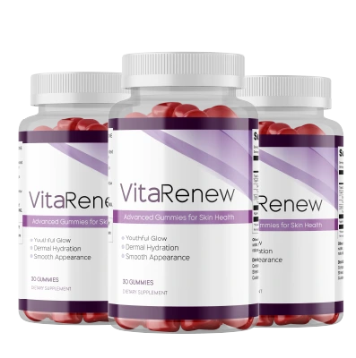
VitaRenew
Vitamin C, copper, zinc, vitamin E, selenium and beta-carotene — the cofactor combination the body's collagen synthesis process depends on, in one daily gummy.
Most readers start with the 2-bottle supply — 60 days is the minimum window for a fair assessment of any internal skin support.
60-day money-back guarantee · One-time purchase, no subscriptionWhy topical products have a structural ceiling at both elbow zones
The skincare industry addresses crepey skin on the arms with a consistent set of topical recommendations: firming body creams with retinol, hyaluronic acid sprays, caffeine-based toning formulas, and products listing collagen or elastin among their ingredients. For the general arm skin these products are consistently used, and for surface hydration they provide a genuine benefit. Surface hydration reduces the visual prominence of crepey texture by improving light reflectance and skin pliability.
The structural ceiling of topical products is determined by skin biology, not by formulation effort. The stratum corneum — the outer protective barrier of the skin — limits molecular penetration based on molecular weight and lipophilicity. The threshold for meaningful transdermal delivery is approximately 500 daltons for small molecules. Collagen has a molecular weight in the hundreds of thousands of daltons; applied collagen does not penetrate the skin surface. Elastin behaves similarly. Even retinoids, the category with the most robust evidence for sub-surface biological activity, primarily affect the dermal-epidermal junction rather than the deep dermis where the collagen scaffold is built.
At the antecubital fossa, the crepey texture reflects mechanical fatigue of collagen fibres in the mid-dermis — a process driven from inside the tissue outward. At the olecranon cap, it reflects both oxidative degradation of fibres and reduction in the dermal support structure beneath the skin. Neither failure mechanism originates at the skin surface, and neither can be addressed by molecules that cannot penetrate beyond the surface.
The enzymatic processes that build and cross-link the dermal collagen scaffold are intracellular processes in fibroblasts. Their cofactors — vitamin C, copper, zinc — are delivered through the bloodstream to the fibroblast. The antioxidant protection of existing fibres operates through cellular mechanisms that similarly depend on systemically available nutrients. The skin surface is the wrong address for these processes.
The formulation standard the biology requires
Against the biology: a working checklist
- Vitamin C at a functional dose. Prolyl and lysyl hydroxylase require consistent adequate supply to maintain hydroxylation rates in active fibroblasts. Trace inclusion at the bottom of a long ingredient list does not constitute functional supply.
- Copper, alongside zinc. Most skin supplements include zinc and omit copper — addressing MMP regulation while missing the cross-linking step where the scaffold actually acquires load-bearing structure. The omission is common and has direct structural consequences at mechanically stressed sites.
- Both vitamin E and selenium, together. The antioxidant pair consistently cited together in dermal oxidative-stress research. At the olecranon cap, where UV exposure means photoaging compounds the intrinsic process year-round, the protective mechanism for existing fibres is more critical than at covered sites.
- Verified manufacturing standards. FDA-registered facility, cGMP certification. Documentable quality assurance, not marketing language.
- Guarantee length matched to the biology. Dermal collagen remodelling is measured at eight to twelve weeks in clinical research. A 60-day guarantee is the minimum that reflects this timeline honestly.
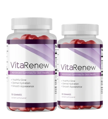
VitaRenew
Of the formulas assessed against this checklist, VitaRenew maps most directly to the dermal cofactor literature. It supplies vitamin C, zinc, copper, vitamin E, selenium, and beta-carotene in a single daily gummy — covering both the enzymatic requirements of collagen synthesis and the antioxidant-protection requirements for existing fibres.
For the elbow specifically, the complete cofactor profile is more relevant than at most other sites. The antecubital fossa requires the collagen synthesis chain to be fully supported — vitamin C for hydroxylation, copper for cross-linking, zinc for turnover regulation — because the mechanical demand on the scaffold there is among the highest of any skin site. The olecranon cap requires the antioxidant mechanism to be complete — vitamin E and selenium together — because UV exposure means the protective function for existing fibres carries more load than at sun-protected sites.
VitaRenew is manufactured in an FDA-registered, cGMP-certified facility, sold as a one-time purchase with no subscription, and backed by a 60-day money-back guarantee. The 2-bottle option provides the minimum 60-day window the dermal biology requires for a meaningful internal assessment.
VitaRenew is a supplement. It supports healthy skin, hair, and nails. It is not intended to diagnose, treat, or cure any condition, and these statements have not been evaluated by the FDA.
On timelines and realistic expectations
The structural changes at the elbow — both the mechanical fatigue history of the antecubital fossa and the photoaging history of the olecranon cap — have accumulated over years or decades. No internal supplement reverses that structural history. What the cofactor research supports is that adequate supply of the molecules the collagen synthesis process requires may help maintain the ongoing rate of that process, and that the antioxidant mechanism may reduce the rate of ongoing oxidative degradation of existing fibres.
Surface texture changes related to hydration and skin barrier function can shift in four to six weeks. Changes in the deeper dermal scaffold are measured at eight to twelve weeks in clinical dermatology research. The mechanical-demand environment at the inner elbow means that any improvement in the scaffold's structural quality will show most clearly in how the skin at the crease behaves under repeated flexion — whether the accordion fold is less prominent when the arm is extended, and whether the texture at the crease after bending returns to shape more quickly.
"The skin at the inside of my elbow had developed this fold that stayed visible even when my arm was straight — I first noticed it properly around 43. I used a couple of firming arm lotions over several months with no real change. After about ten weeks on VitaRenew the fold at the inner crease is noticeably less prominent and the skin above it doesn't look as loose. The outer elbow also looks less ashy. I wasn't expecting the outer elbow to change but it has."
— Joanna W., 45, verified purchaser
"I kept seeing the inside of my elbow in video calls — the crinkle when I rested my chin on my hand. I'd never paid attention to elbow skin before but once I saw it I couldn't unsee it. I started VitaRenew in February and by early April I noticed the texture inside the crease and on the outside of the elbow both looked different — more like arm skin used to look. The accordion fold is still there but much less deep."
— Patricia H., 48, verified purchaser
Individual results vary. These testimonials represent personal experiences and may not reflect typical outcomes.
On asymmetric or atypical changes: The structural changes described in this article are gradual and bilateral. If you notice changes at the elbow that are asymmetric, rapid, or accompanied by joint pain, swelling, redness, or warmth in the joint itself, those warrant clinical evaluation before any supplement consideration. Skin texture changes at the elbow that do not follow the gradual, bilateral pattern should be assessed by a healthcare professional.
The short version
Crepey skin at the elbows develops through two distinct structural pathways. The antecubital fossa — the inner crease — accumulates mechanical fatigue from the highest flexion-deformation cycle rate of any skin site on the upper limb, compounded by the decline in collagen synthesis that begins in the mid-twenties. The olecranon cap — the outer elbow — combines an absent subcutaneous fat foundation, chronic friction from hard surfaces, and decades of UV exposure that layers photoaging onto intrinsic aging in a way that covered arm skin does not experience.
Both failure mechanisms ultimately trace back to the same collagen maintenance process: fibroblast-mediated synthesis of cross-linked, load-bearing dermal scaffold, dependent on enzymatic cofactors delivered through the bloodstream. Vitamin C for the hydroxylation steps, copper for the cross-linking step, zinc for turnover regulation, and vitamin E and selenium together for antioxidant protection of existing fibres — particularly relevant at the UV-exposed olecranon cap. Topical products cannot reach these processes. Internal cofactor supply addresses the mechanism at its actual address.
Product referenced above
VitaRenew
The cofactor-first formula for skin, hair and nails. FDA-registered, GMP-certified facility. No subscription. Ships from the US.
Start with the 2-bottle supply — 60 days gives the biology time to show a real response.
60-day money-back guarantee · No subscription · Ships from the USBackground Reading
- Pullar JM, Carr AC, Vissers MCM. "The Roles of Vitamin C in Skin Health." Nutrients, 2017.
- Rucker RB et al. "Copper, lysyl oxidase, and extracellular matrix protein cross-linking." American Journal of Clinical Nutrition, 1998.
- Varani J et al. "Decreased collagen production in chronologically aged skin." American Journal of Pathology, 2006.
- Uitto J. "The role of elastin and collagen in cutaneous aging." Journal of Drugs in Dermatology, 2008.
- Yaar M, Gilchrest BA. "Photoageing: mechanism, prevention and therapy." British Journal of Dermatology, 2007.
- Ogawa Y et al. "Zinc and skin disorders." Nutrients, 2018.
- Brennan M et al. "Protein crosslinking in aging skin." Journal of Investigative Dermatology, 2003.