Body Skin · Research Explainer
Why the Skin on Your Knees Looks Crepey After 40
Of all the places crepey skin develops in midlife, the knees surprise women the most — precisely because the biology there is unlike any other site on the body. Three structural vulnerabilities converge at the knee, and nowhere else, making it one of the most predictable places for intrinsic aging to show visibly after 40.

The question of why knee skin develops crepey texture is one that tends to arrive in the summer months, when shorts and swimwear bring the area into view after months of coverage. Women notice it above the knee — where the skin seems to have loosened and folded — and around the kneecap itself, where a papery, crinkled texture appears when the leg is extended. Some notice it behind the knee in the crease, where the skin gathers when the leg bends.
The consistency of that pattern is not random. The knee is, structurally, one of the most demanding sites in the body for skin. The set of pressures it faces — biomechanical, hormonal, and environmental — is unlike those at the thighs, the arms, or the face. Understanding why the knee develops this pattern requires looking at three factors that converge there and nowhere else at the same intensity.
The three structural vulnerabilities of knee skin
First: the subcutaneous fat pad diminishes here with age in a way that directly exposes the skin. The skin over the patella and the area just above it sits on a layer of subcutaneous fat that provides physical padding and volume beneath the skin's surface. This fat pad is thinner at the knee than at the thighs or buttocks to begin with — the knee is a bony prominence, not a soft-tissue region — and it responds to the changes of midlife more visibly than padded areas. As the fat pad reduces with age, the skin over the patella loses its underlying support. Instead of sitting flush against a rounded, supported surface, it begins to drape. The loose quality above the knee — where the skin seems to have stretched and not returned — is in large part the dermis settling over a reduced support structure.
Second: the knee undergoes more mechanical deformation cycles per day than almost any other joint. Every step involves flexion and extension of the knee. Sitting, standing, climbing stairs — the motion range is substantial, and the skin over the anterior and superior knee participates in every cycle. Skin is viscoelastic: it stretches and recovers. But that recovery depends on intact, well-organised collagen and elastin fibres in the dermis. Over decades, the accumulated mechanical fatigue of hundreds of thousands of flex-extend cycles degrades the organised cross-linked structure of those fibres. The collagen network that once returned the skin to its resting shape begins to lose that capacity. Add the decline in collagen synthesis that begins in the mid-twenties at roughly one percent per year, and the mechanical demand on the dermis progressively outpaces its repair capacity. The result is skin that stretches under load and does not fully return — the defining characteristic of crepey texture.
Third: the knee is one of the most UV-exposed sites below the neck. Unlike the inner thighs, which spend most of the year covered, the knees and the skin above them are regularly exposed to UV radiation — through shorts, swimwear, skirts, and outdoor activity over decades. This means that in addition to the intrinsic aging affecting all skin equally, the knee also carries the photoaging load that accelerates the breakdown of collagen and elastin fibres through UV-generated reactive oxygen species and MMP enzyme activation. The two processes — intrinsic aging and photoaging — run in parallel at the knee in a way they do not at covered sites.
Fat pad reduction that exposes the dermis, the highest mechanical load cycle of any joint in daily ambulation, and regular UV exposure that adds photoaging to the intrinsic process. The knee is the only site below the neck where all three converge. The biology makes the pattern entirely predictable.
The role of estrogen in accelerating the timeline
The three structural vulnerabilities above are not new at 40 — they have been accumulating since early adulthood. What changes in the early-to-mid forties, for most women, is the hormonal environment that has been partially compensating for that structural decline.
Estrogen supports fibroblast activity in the dermis — the cells responsible for synthesising and maintaining collagen and elastin. It helps regulate hyaluronic acid production, which maintains the physical plumpness of the dermal matrix. It also has a role in sebaceous gland function, affecting the baseline hydration of the skin surface. When estrogen begins declining during perimenopause, the rate of structural change in the dermis accelerates across the whole body. But it accelerates most visibly in sites that were already under structural stress — which is why the knees, already compromised by fat pad reduction and mechanical fatigue, often show the change so sharply in the early-to-mid forties.
Estrogen decline does not cause crepey skin at the knee. What it does is remove a layer of hormonal support that had been partially compensating for the structural vulnerabilities that were always there. The change feels sudden — and often is, in terms of visible texture — but the underlying structural decline was gradual.
What is happening at the molecular level in the dermis
The visible crepey texture at the knee reflects a specific degradation in the organisation of the dermal scaffold. The dermis is built around a cross-linked mesh of collagen fibres and elastin fibres, embedded in a hydrating matrix of hyaluronic acid and proteoglycans. This scaffold provides both the physical volume of the skin and its elastic recovery — its ability to return to shape after deformation.
The scaffold is maintained by fibroblasts, which continuously produce, organise, and turn over its protein components. This maintenance process requires specific enzymatic steps, each of which has molecular cofactor requirements:
The collagen triple helix must be hydroxylated at proline and lysine residues before it achieves structural stability. This hydroxylation is performed by prolyl hydroxylase and lysyl hydroxylase — enzymes that require vitamin C as an obligate cofactor. Without adequate vitamin C, the procollagen strands produced are structurally incomplete. They cannot form proper triple helices. The result is that fibroblast activity, even when maintained, does not produce functional collagen if the cofactor supply is insufficient.
Individual collagen and elastin strands must then be cross-linked into a load-bearing network by lysyl oxidase. This step — which converts protein strands into structural scaffold — requires copper as its obligate cofactor. Without copper, fibres are synthesised but remain as disorganised strands rather than integrated scaffold. The cross-linking step is where the dermis actually becomes structural tissue capable of withstanding mechanical load. Copper is consistently underrepresented in commercial skin supplements despite appearing in the peer-reviewed dermal biology literature as centrally as vitamin C.
Zinc regulates the matrix metalloproteinases that govern collagen turnover and participates in the wound-signalling cascade that maintains repair capacity in the dermis. Its role is supportive throughout the synthesis and maintenance cycle.
A parallel mechanism involves oxidative degradation of existing fibres. Reactive oxygen species — generated by UV exposure, metabolic activity, and inflammation — fragment collagen and elastin in the dermis. Vitamin E and selenium address this mechanism through complementary antioxidant pathways consistently cited together in dermal oxidative-stress research. Their role is protective rather than synthetic: sustaining the longevity of existing fibres while the synthesis pathway works to replace them.
Referenced in this article
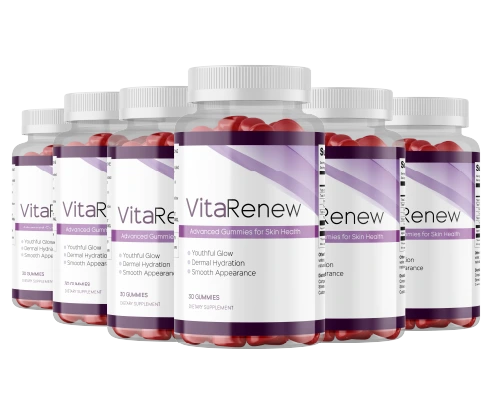
VitaRenew
Vitamin C, copper, zinc, vitamin E, selenium and beta-carotene — the cofactor combination the body's collagen synthesis process depends on, in one daily gummy.
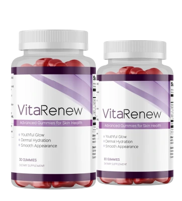
Most readers start with the 2-bottle supply — 60 days is the minimum window for a fair assessment of any internal skin support.
60-day money-back guarantee · One-time purchase, no subscriptionWhy topical approaches have a structural ceiling at the knee
The market for knee-area firming products is substantial, and several categories of product are commonly used. Firming body creams with collagen or elastin on the label, retinol body lotions, caffeine-based formulas, and hyaluronic acid sprays are all widely recommended for crepey skin on and above the knee.
The benefit these products offer is real but limited. Good hydration of the skin surface reduces the visual prominence of fine surface texture. A well-moisturised skin surface reflects light more evenly and is more pliable, which makes crepey texture less pronounced. That effect is genuine and worth maintaining as part of a daily routine.
The structural ceiling is determined by skin biology. The stratum corneum — the outer protective barrier of the skin — restricts molecular penetration based on molecular weight. Molecules above approximately 500 daltons do not penetrate in meaningful quantities. Collagen is far above that threshold; applied collagen remains at the surface. Topical elastin does the same. Even retinoids, which have the most robust evidence of any topical ingredient for sub-surface activity, affect the dermal-epidermal junction — not the deep dermis where the collagen scaffold is built and maintained.
The crepey texture visible on the knees reflects a failure of internal scaffold maintenance. The enzymatic machinery that builds and cross-links that scaffold requires cofactors that are delivered through the bloodstream, not through the skin surface. Topical products cannot reach the process that generates the structural problem.
What the research supports for internal cofactor supplementation
Against the biology, a working checklist
- Vitamin C at a functional dose. Prolyl and lysyl hydroxylase require consistent adequate supply to maintain hydroxylation rates in active dermis. A trace inclusion does not constitute functional supply.
- Copper, alongside zinc. Most skin supplements include zinc and omit copper — which means they address collagen turnover regulation while missing the cross-linking step where scaffold structure forms. The omission is common and consequential.
- Both vitamin E and selenium, together. The antioxidant pair cited together in dermal oxidative-stress research. At the knee, where UV exposure adds photoaging to the intrinsic process, the protective mechanism for existing fibres is more load-bearing than at covered sites.
- Verified manufacturing standards. FDA-registered facility, cGMP certification. Documentable facts, not marketing language.
- A guarantee length that matches the biology. Dermal collagen remodelling is measured at eight to twelve weeks in clinical research. A 60-day guarantee is the minimum that reflects this timeline honestly.
VitaRenew
Of the formulas assessed against this checklist, VitaRenew maps most directly to what the dermal cofactor literature describes. It supplies vitamin C, zinc, copper, vitamin E, selenium, and beta-carotene in a single daily gummy — covering both the enzymatic requirements of collagen synthesis and the antioxidant-protection requirements for existing fibres. At the knee specifically, where UV exposure means photoaging compounds the intrinsic process, the antioxidant component (vitamin E and selenium together) is particularly relevant.
VitaRenew is manufactured in an FDA-registered, cGMP-certified facility, sold as a one-time purchase with no subscription, and backed by a 60-day money-back guarantee. The 2-bottle option provides the 60-day minimum window the dermal biology requires for a meaningful internal assessment.
VitaRenew is a supplement. It supports healthy skin, hair, and nails. It is not intended to diagnose, treat, or cure any condition, and these statements have not been evaluated by the FDA.
On realistic expectations and timelines
The structural changes that crepey skin at the knee reflects have accumulated over years or decades. An internal supplement does not reverse structural history. What the cofactor research supports is that adequate supply of the molecules the collagen synthesis process requires may help maintain the ongoing rate of that process — and that the protective antioxidant mechanism may reduce the rate of ongoing degradation of existing fibres.
Surface texture and hydration can shift in four to six weeks with internal support. Changes in the deeper dermal scaffold are measured at eight to twelve weeks in clinical dermatology research. Individual variation — in hormonal status, UV history, baseline nutrition, genetics, and starting point — is real and substantial. Two women using the same formula for the same duration may have meaningfully different experiences, both within the normal range of variation.
"I noticed the crepey texture above my knees around 44 — I could see it clearly when I sat with my legs out. I had tried a couple of firming creams without much change. After about nine weeks on VitaRenew I noticed the quality of the skin above the knee looked different in photos — smoother and less folded. The kneecap area also looks less papery. It's a real difference that has held over four months."
— Denise K., 46, verified purchaser
"The skin around my knees was the thing I noticed first, in a photo from a family event. I'm a runner, so I was used to seeing my legs in shorts, and the change surprised me. I started VitaRenew in April and by early June the texture around and above the knees was noticeably smoother. The crepe paper look when I extended my legs is much less pronounced. I'm continuing with the 3-bottle supply."
— Rebecca T., 50, verified purchaser
Individual results vary. These testimonials represent personal experiences and may not reflect typical outcomes.
On asymmetric or rapid change: the structural changes described in this article are gradual and bilateral. If you notice changes at the knee that are asymmetric, rapid, or accompanied by swelling, pain, redness, or warmth in the joint or surrounding skin, those warrant clinical evaluation before any supplement response. Changes outside the pattern of gradual bilateral skin texture shift should be assessed by a healthcare professional first.
The short version
Crepey skin on and above the knees after 40 results from the convergence of three structural factors found at no other site below the neck: progressive reduction of the subcutaneous fat pad that supports the skin over the patella, the highest mechanical deformation load of any joint in daily movement, and regular UV exposure that layers photoaging onto the intrinsic process. Estrogen decline in perimenopause accelerates the visible result of structural vulnerabilities that have been accumulating for decades.
Surface products hydrate and temporarily reduce the visual prominence of fine texture — a real benefit worth maintaining. The collagen scaffold beneath the surface depends on enzymatic machinery that requires specific cofactors delivered through the bloodstream. Those cofactors — most centrally vitamin C, copper, and zinc, with vitamin E and selenium for antioxidant protection — are distinct from dietary protein and commonly absent or underdosed in standard skin products. At the knee specifically, where photoaging compounds the intrinsic process, the complete cofactor picture matters more than at purely covered sites.
Product referenced above
VitaRenew
The cofactor-first formula for skin, hair and nails. FDA-registered, GMP-certified facility. No subscription. Ships from the US.
Start with the 2-bottle supply — 60 days gives the biology time to show a real response.
60-day money-back guarantee · No subscription · Ships from the USBackground Reading
- Pullar JM, Carr AC, Vissers MCM. "The Roles of Vitamin C in Skin Health." Nutrients, 2017.
- Rucker RB et al. "Copper, lysyl oxidase, and extracellular matrix protein cross-linking." American Journal of Clinical Nutrition, 1998.
- Thornton MJ. "Estrogens and aging skin." Dermato-Endocrinology, 2013.
- Varani J et al. "Decreased collagen production in chronologically aged skin." American Journal of Pathology, 2006.
- Uitto J. "The role of elastin and collagen in cutaneous aging." Journal of Drugs in Dermatology, 2008.
- Ogawa Y et al. "Zinc and skin disorders." Nutrients, 2018.
- Yaar M, Gilchrest BA. "Photoageing: mechanism, prevention and therapy." British Journal of Dermatology, 2007.